Overview
Radboudumc (Radboud University Medical Center) is located in Nijmegen, Netherlands and is one of the leading academic hospitals and research institutes, affiliated with Radboud University.
Professor Sommerdijk is a leading pioneer in biomineralization, nanoscale materials, and advanced electron microscopy. He established the Radboud Electron Microscopy Center that supports the advanced electron microscopic analysis of biological materials using some of the most advanced imaging and analysis strategies.
The special focus areas are 3D volume imaging, correlative light and electron microscopy, and cryo-electron microscopy. Through a recently awarded NWO-Groot project they now also serve as a National Facility for liquid phase EM of biological materials (BIOMATEM).
The Challenge
After hearing about compressed sensing from Professor Nigel Browning, CSO at SenseAI, Prof Sommerdijk investigated the benefits of SenseAI electron microscopy imaging for bone formation processes and in particular the layering and mineralisation of collagen and apatite.
Dr Luco Rutten is a post-doctoral researcher in Prof Sommerdijk’s group and specialises in the sparse imaging of bio materials.
He said: “When sampling biological materials, TEM works by analysing very thin samples, where you need to work with low electron doses so as not to damage the samples. This generates noisier images at lower resolution. At the same time, to study biological process we often need a larger volume to be more physiological relevant. We use STEM for these thicker samples where we can get a larger depth of focus. In situ is the holy grail for both however, particularly when you want a movie of nano-processes and for that you need a very low dose.”
How SenseAI helped
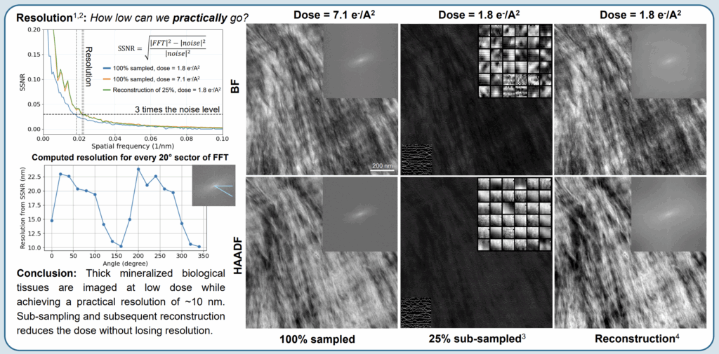
SenseAI has been used by the team at Radboudumc to generate high-resolution images, using a much lower dose. In recent work with collagen fibrils, SenseAI generated the same quality image as a full-dose image, but with just a fraction of the electron dose.
Dr Rutten said: “We have validated the performance of SenseAI and it has exactly the same resolution, using just a quarter of the dose. This supercharges our research as well as speeding up the experiments by 4x”.
“We have also used SenseAI for full-sample TEM denoising in 3D as a post-processing tool and the dictionary learning and inpainting are able to significantly improve the quality and usability of the images”

Future applications
Professor Sommerdijk is looking to build on his research with even more advanced biological structures and processes. Some of which have more contrast such as membrane fusion but others have even more challenging tissues such as the brain i.e. delivery of neurotransmitters in synaptic clefts.
He said: “Our goal is to look at cells with electron doses of less than 0.05 electrons per square Angstrom as this is the limit of when enzymes are active. This is where we can use sparse sampling to go to new levels. To reach this limit we need to combine sparse imaging with other strategies to reduce the electron damage e.g. graphene protection. We foresee great potential for the in situ imaging for biological processes using these methods.
We see potential in other techniques where SenseAI can potentially power us forward even more – Cryo FIB SEM, Raman Spectroscopy and Confocal Microscopy. SenseAI enabled us to break through technical barriers to take our research to new levels. It is also straightforward and quick to use. We are excited about our next leg of the journey”


