Why fast image capture in Electron Microscopy is so important and how can we image in real time
The speed at which images are acquired – the acquisition speed – has a huge impact on the efficiency, and capability of Electron Microscopy users the world over.
Why is Acquisition Speed so important?
In a nutshell, the whole usage of Electron Microscopy hangs off acquisition speeds. It’s the key to unlocking key future advancements in imaging and analysis. The faster users can obtain images, more and more benefits open up such as:
- Throughput and productivity
- Capturing dynamic processes/analysis
- Reducing drift and artefacts
- Improving image quality
- Reducing cost
How SenseAI is speeding up Acquisition Speeds
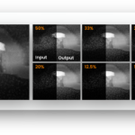
Using proprietary sub-sampling methodologies, SenseAI can generate the same high-quality images and video feeds with up to 100x less data (1% of the original data). This can massively speed up the imaging process. At its core, SenseAI utilises an ultra-fast implementation of a blind inpainting algorithm – the beta process factor analysis (BPFA) algorithm. BPFA is used to learn a sparse representation of a dataset, which is then used to reconstruct that entire dataset. Blind dictionary learning and inpainting algorithms allow dictionaries to be learned and images to be inpainted directly from subsampled data – no external training data is required
These methods have been demonstrated to work on many scanned imaging techniques, such as SEM and STEM, and even 4D-STEM where it can effectively turn the acquisition into a live i.e. real-time process rather than one that takes minutes or sometimes hours to acquire each image.
Unlocking speed and real-time, live imaging in 4D STEM
The groundbreaking advantage of SenseAI for experiments is that the ability to create live reconstructions using subsampled data in 4D STEM which enables real time adjustments. While you’re on the microscope, we can change the magnification, alignment, or quickly pan to find a/the region of interest, and we can optimise our analysis to get the best image. This is revolutionising Electron Microscopy imaging and enabling analysis that we’ve simply not been able to do previously.
Noise doesn’t have to be a problem
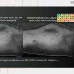
SenseAI is also a powerful denoiser tool. Faster imaging effectively reduces signal-to-noise and increased noise, due to reduced measuring times. Using SenseAI as a denoiser is simple to use and works across all techniques – 4D-STEM, 2D-STEM, TEM, and SEM. It can be used in post-processing (i.e. to denoise any images acquired by any other methods) or the entire workflows (i.e. for acquisition as well).
Examples of SenseAI speeding up acquisition times
SenseAI is speeding up acquisition times for labs, academia and industry the world over. Here’s a quick glimpse into some of the speed improvements being achieved:
Dr Luco Rutten, Radboud University Medical Center says: “We have validated the performance of SenseAI and it has exactly the same resolution, using just a quarter of the dose. This supercharges our research as well as speeding up the experiments by 4x.”
Dr Giuseppe Nicotra, Head of Sub-Ångstrom Electron Microscope LAB at CNR-IMM adds: “With SenseAI we are able to image in real time. There’s nothing worse than when you acquire a 4D STEM data set and then you’re unable to know how good it is until you’ve gone away, processed and analysed it, and have to re-acquire the datasets.”
And finally Professor Roland Fleck, Director of Centre for Ultrastructural Imaging at Kings College London says: “One of the main advantages of using SenseAI is if I’m only collecting, say, 20 or 25% of the field of view by using the compressed sensing technology, I’m essentially working four or five times faster than I can otherwise. So, the time required to complete an entire volume is significantly reduced, which is less stressful on the instrument, less stressful on the operator, and generates much higher success rates.”