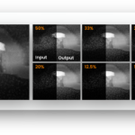


4D STEM nADF Negative Annular Dark Field
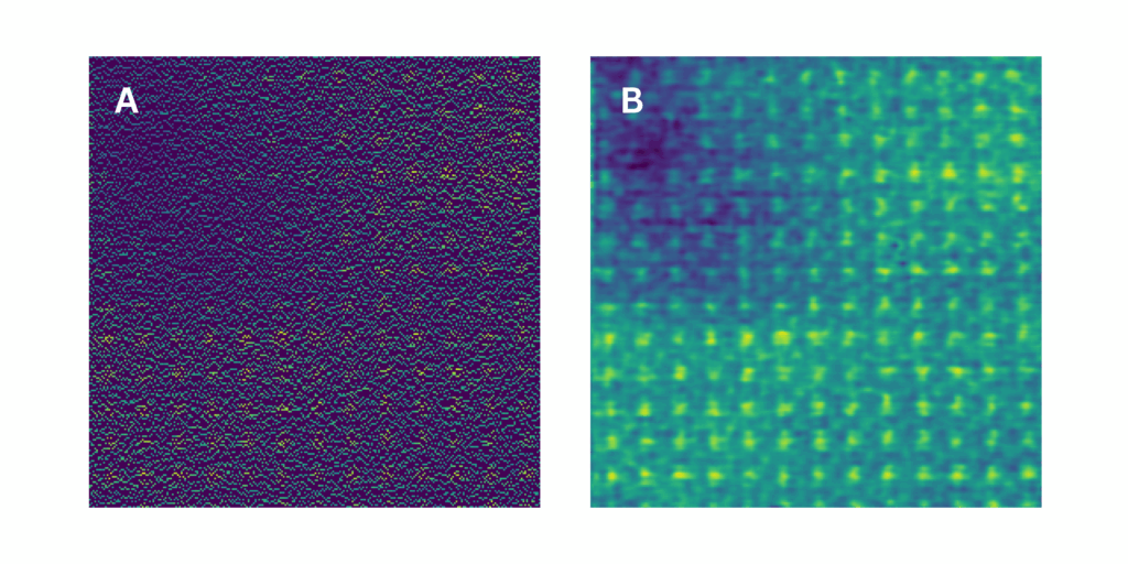
Left (A): Subsampled layer using 25% of original data points. Right (B): Reconstruction using SenseAI. Total scan time is approximately 0.635 seconds.
Generated from one 4D-STEM data set acquired by SenseAI Vision subsampling and inpainting software using the new JEOL GrandARM2 at the University of Liverpool and using a Direct Electron CeleritasXS camera by Dr. Mounib Bahri and Dr Alex W. Robinson. The sample is [100] Strontium Titanate SrTiO3 prepared by FIB.
We’re delighted to announced the launch of our new 4D STEM product. The software enables microscopists to perform 4D STEM live, with significantly less beam exposure and using less than 10% of the data.
4D Scanning Transmission Electron Microscopy (4D-STEM) has long been regarded as one of the most powerful but challenging techniques in electron microscopy. Historically, its adoption and success has been limited by complex workflows, instability (drift), beam damage, slow frame rates, vast data sizes, and a lack of pre-acquisition surveying.
SenseAI’s 4D STEM imaging solution changes this paradigm by dramatically streamlining the entire process—from alignment through to analysis. Users can dynamically image and analyse areas of interest in real time just like they would expect to with conventional STEM, all whilst using less than 10% of the data.
SenseAI also images at previously unachievably low doses to preserve sample integrity, enabling high-quality data collection even on the most sensitive samples.
Professor Nigel Browning, Chair of Electron Microscopy at Liverpool University and CSO of SenseAI says: “Now microscopists the world over, using their existing hardware, can perform research they simply couldn’t do before.
“Traditionally slow acquisition times means that only the most stable microscopes could consider using 4D STEM. Because of SenseAI’s faster imaging via the use of subsampling, stability issues are minimised and high quality data are maintained. It is the pinnacle of electron microscopy but now simplified and accessible by all.”
Giuseppe Nicotra, Head of Sub-Ångstrom Electron Microscope LAB at CNR-IMM who is using SenseAI for 4D STEM research says: “There’s nothing worse when you acquire a 4D STEM data set and then you’re unable to know how good it is until you’ve processed and analysed it, and then have to re-acquire the datasets. This is a lengthy process consuming huge amounts of data. With SenseAI you can see 4D STEM images live and make adjustments on the fly.
“Many of our specimens are also very beam sensitive. With SenseAI we can work at previously unachievably low doses to preserve sample integrity and benefit from superior data.
“Electron microscopy datasets are huge, even generating these large datasets requires very long acquisition times. In our experiments using SenseAI, we are able to subsample using just 10% or less of the original data. Maintaining large data volumes in a data centre is costly but we can now preserve and easily handle all of the required information using a lot less storage.”
The 4D STEM software can also support live virtual detectors, live DPC, live centre of mass and post processing techniques such as ptychography.